A cerebral aneurysm isn't something most people hear about until it's too late. It's a weak spot in a blood vessel in the brain that balloons out like a tiny blister. Most of the time, it doesn't cause symptoms. But when it ruptures, it can trigger a life-threatening bleed called a subarachnoid hemorrhage. About 3.2% of people worldwide have one of these unruptured aneurysms, and each year, roughly 9 to 10 out of every 100,000 people experience a rupture. The death rate within the first day is between 30% and 40%. That’s why understanding the risks and treatment options matters - whether you’ve been diagnosed or just want to know what to watch for.
What Makes an Aneurysm Likely to Burst?
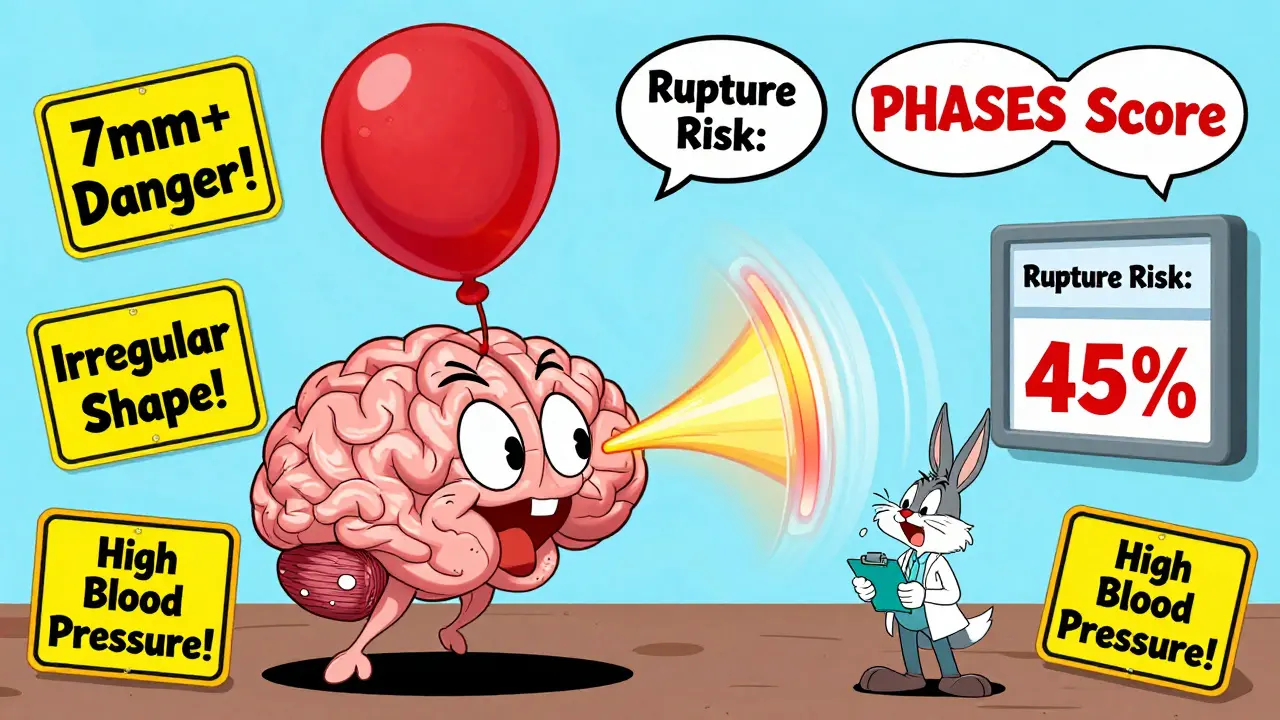
Not all aneurysms rupture. Many stay small and never cause trouble. But some do - and doctors use a mix of factors to guess which ones might. Size is the biggest red flag. Aneurysms larger than 7 millimeters are over three times more likely to burst than smaller ones. Shape matters too. If the bulge looks irregular, has a daughter sac (a smaller bulge branching off), or is asymmetrical, the risk jumps. Aneurysms with these features have nearly a 3-fold higher rupture risk compared to smooth, round ones.
Location plays a huge role. Aneurysms at the anterior communicating artery (AComm) are especially dangerous. Even if they’re small - under 5 mm - they can still rupture. Studies show these are 2.4 times more likely to burst than aneurysms in other areas. Middle cerebral artery aneurysms also carry high risk. In contrast, aneurysms in the back of the brain (posterior circulation) are rarer but harder to treat if they do rupture.
Then there’s blood flow. Advanced imaging and computer modeling show that aneurysms with low or chaotic blood flow along their walls are far more likely to rupture. This kind of flow stresses the vessel wall over time, breaking down its structure. About 83% of ruptured aneurysms show this pattern, while only 42% of unruptured ones do.
Who’s at Higher Risk?
Some risks you can’t change. Age is one. People over 65 have a 2.7 times higher risk of rupture than younger adults. Women are more likely to develop aneurysms than men - about 1.6 times more often. And if two or more close family members have had one, your risk jumps fourfold.
But there are things you can control. High blood pressure is the biggest modifiable risk. If your systolic pressure stays above 140 mmHg, your rupture risk increases by 2.3 times. Smoking is even worse. Current smokers face a 3.1 times higher risk than non-smokers. And it’s not just about smoking - the more you smoke, the higher the risk. People who smoke 10 or more cigarettes a day see their risk rise by 47%. Heavy drinking adds to the danger. More than 14 drinks per week increases rupture risk by 32%.
Biological factors are also at play. Studies show higher levels of inflammation markers like IL-6 and CRP in people who’ve had ruptures - up to 37% and 29% higher respectively. Genetics play a role too. Certain gene variants linked to collagen strength and immune response make some people more vulnerable. The HUNT study found 17 genetic locations tied to aneurysm formation and rupture.
The PHASES and ELAPSS Scores: Predicting the Future
Doctors don’t guess - they use tools. The PHASES score is the most widely used. It combines six factors: your population’s baseline risk, whether you have high blood pressure, your age, the size of the aneurysm, whether you’ve had a previous bleed, and its location. Each factor adds points. A score of 0-3 means a 3% chance of rupture in five years. A score of 9-10 means a 45% chance. That’s a massive difference.
Each point increase in PHASES raises the risk by 32%. So if your score is 6 or higher, most experts recommend treatment. For lower scores, monitoring with yearly MRI or MRA scans is often enough.
Another tool, the ELAPSS score, focuses on five factors: earlier subarachnoid hemorrhage, location, age, previous SAH, size, and shape. It gives you a 1-year rupture risk estimate. The newer triple-S model - size, site, shape - is especially useful after an aneurysm has been seen to grow. It can predict risk over 6 months, 1 year, or 2 years with good accuracy.
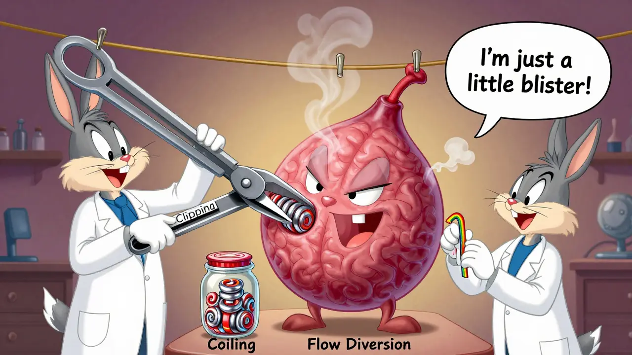
Treatment Options: Clipping, Coiling, and Flow Diversion
If treatment is needed, there are three main approaches. The oldest is microsurgical clipping. A neurosurgeon opens the skull, finds the aneurysm, and places a tiny titanium clip across its neck. This stops blood from flowing into it. It’s been done since 1937. Success rates are high - 95% of aneurysms are completely sealed off. Long-term cure rates are 88-92%.
The second option is endovascular coiling. A catheter is threaded up from the groin into the brain. Platinum coils are then pushed into the aneurysm. They cause a clot to form inside, sealing it off. First done in 1991, it’s now the most common treatment. At six months, 78-85% of aneurysms are fully blocked. It’s less invasive than surgery, so recovery is faster. But it has a catch: 15.7% of coiled aneurysms need another procedure later, compared to just 6.2% for clipped ones.
The third option is flow diversion. This uses a special stent - like the Pipeline Embolization Device - placed in the artery. It redirects blood away from the aneurysm, letting it slowly clot and shrink. It’s especially good for large or wide-necked aneurysms. The PED-PLATINIUM trial showed 85.5% complete occlusion at one year. The WEB device, approved in 2019, works well for aneurysms at artery branches. It achieved 71.4% occlusion at one year in the WEBCAST trial.
Which Treatment Is Right for You?
There’s no one-size-fits-all answer. It depends on the aneurysm - and you.
- Size and shape: Wide-necked aneurysms (>4 mm) often need flow diversion. Small, narrow ones can be coiled.
- Location: Aneurysms in the back of the brain (posterior circulation) have a 22% higher complication risk with clipping. Coiling or flow diversion are often preferred.
- Age: Patients over 70 have a 35% higher risk of surgical complications. Less invasive options are safer here.
- Health: If you have uncontrolled high blood pressure, your risk during any procedure goes up by 28%.
The ISAT trial showed coiling reduced 1-year death risk by 22.6% compared to clipping (8.1% vs 10.1%). But over 12 years, the survival difference evened out. Quality of life after coiling was better - EQ-5D scores were 0.82 at one year, compared to 0.76 for surgery.

What About Not Treating?
Many small aneurysms are watched, not treated. The UCAS Japan study found that unruptured aneurysms under 5 mm have a 0.2% to 0.7% 5-year rupture risk - depending on location. For someone with no other risk factors, that’s very low. Annual MRA scans are usually enough. But if the aneurysm grows, even slightly, the risk spikes. That’s why follow-up imaging is critical.
Medical management is key for everyone. Lowering blood pressure to under 130/80 mmHg reduces rupture risk. Quitting smoking cuts risk by 54% within two years. Cutting back on alcohol helps too. These aren’t optional - they’re part of the treatment plan.
What Happens After Treatment?
Successful treatment cuts long-term rupture risk dramatically. Without treatment, the chance of another rupture within 10 years is 68%. With successful clipping or coiling, that drops to 2.3%.
Complications vary. Clipping has a 4.7% permanent morbidity rate and 1.5% mortality. Coiling is slightly safer: 3.9% morbidity and 1.1% mortality. Flow diversion has a 5.2% morbidity rate, but only 0.8% mortality - making it a strong option for complex cases.
Recovery time differs too. Clipping usually means a hospital stay of 5-7 days and weeks of rest. Coiling often allows patients to go home in 1-2 days. Flow diversion requires blood thinners for months and careful monitoring.
What’s Next in Research?
Science is moving fast. Researchers are now using machine learning to analyze 42 different features of an aneurysm - shape, flow patterns, wall thickness - to predict rupture better than current scores. Genetic testing may soon identify people at highest risk before an aneurysm even forms. New devices are in trials, including bioresorbable stents that dissolve after doing their job.
The goal isn’t just to treat - it’s to prevent. Better screening, smarter risk tools, and less invasive treatments are making cerebral aneurysms less deadly than they used to be.
Can a cerebral aneurysm be detected before it ruptures?
Yes. Many are found accidentally during brain scans done for other reasons, like migraines or head injuries. High-risk individuals - such as those with a family history or certain genetic conditions - may be screened with MRA or CTA scans. Routine screening isn’t recommended for the general public, but if you have two or more close relatives with aneurysms, talk to a neurologist about testing.
What are the warning signs of a ruptured aneurysm?
A ruptured aneurysm often causes a sudden, severe headache - described as the "worst headache of your life." Other signs include nausea, vomiting, stiff neck, blurred vision, sensitivity to light, seizures, or loss of consciousness. If you or someone else experiences this, call emergency services immediately. Time is critical.
Is it safe to fly with an unruptured aneurysm?
For most people with small, unruptured aneurysms, flying is safe. Cabin pressure changes during flight don’t significantly increase rupture risk. However, if your aneurysm is large (>7 mm), irregular, or you’ve recently been diagnosed, consult your neurologist. They may recommend delaying travel until you’ve been evaluated or treated.
Can lifestyle changes reduce rupture risk?
Absolutely. Controlling blood pressure, quitting smoking, and limiting alcohol are the most effective steps. Studies show quitting smoking cuts rupture risk by 54% within two years. Keeping systolic pressure below 130 mmHg reduces strain on vessel walls. Avoiding heavy lifting and extreme stress may also help, though the evidence is less clear. These aren’t just helpful - they’re essential.
How often should I get scanned if I have an unruptured aneurysm?
For small aneurysms (<5 mm) with low PHASES scores, annual MRA scans are standard. If the aneurysm is larger, irregular, or growing, scans may be done every 6 months. If you’ve had treatment, follow-up imaging is needed at 6 months and then yearly to check for recurrence. Always follow your specialist’s advice - timing depends on your individual risk profile.


Kenzie Goode
February 24, 2026 AT 13:31Wow, this post really opened my eyes. I had no idea aneurysms could be so silent until they’re not. The part about shape and blood flow patterns? Mind-blowing. I’ve had migraines for years and never thought to get screened - maybe I should. Thanks for laying this out so clearly.
Dominic Punch
February 24, 2026 AT 22:39Let’s cut through the noise: if you’re over 50, have HTN, and smoke - you’re playing Russian roulette with your brain. Coiling isn’t ‘less invasive’ - it’s the only sane choice for most. Clipping is a 1970s solution with a 2024 price tag. And don’t get me started on people who think ‘I’m fine’ because their aneurysm is under 5mm. Location matters more than size. AComm? That’s a ticking clock. Stop waiting for symptoms. Get scanned.
Valerie Letourneau
February 26, 2026 AT 16:06Thank you for sharing such a comprehensive and meticulously researched overview. The integration of PHASES and ELAPSS scores into clinical decision-making is particularly commendable. I appreciate how the post balances statistical rigor with human relevance - especially the emphasis on modifiable risk factors. In Canada, we’ve seen a marked increase in MRA accessibility over the past decade, and this kind of public education is vital to ensuring equitable outcomes. Well done.
Cory L
February 28, 2026 AT 04:41Bro. I had a cousin who went from ‘mild headache’ to ‘brain bleed’ in 12 hours. No warning. No second chances. The fact that a 7mm bulge triples your odds? That’s not a statistic - that’s a death sentence waiting to happen. And smoking? Dude, it’s not even a choice anymore. If you’re puffin’ and over 40, you’re basically paying rent on a time bomb. Quit. Get checked. Don’t be the guy who says ‘I’ll do it next year.’ Next year doesn’t exist for these things.
Bhaskar Anand
February 28, 2026 AT 19:19Stephen Archbold
March 2, 2026 AT 11:25Just had my first MRA last month - found a 4mm AComm aneurysm. Was terrified. Then I read this. Honestly? This is the clearest breakdown I’ve seen. Flow diversion sounds scary but the 0.8% mortality? That’s better than a tonsillectomy. I’m gonna go with PED. My neurologist said ‘if you’re gonna do it, do it right.’ Thanks for the data. And yeah - I quit smoking yesterday. No more excuses.
Timothy Haroutunian
March 3, 2026 AT 06:56Look. I’ve read like 17 papers on this. The entire field is built on shaky assumptions. PHASES? It’s based on 2008 data. Most studies use retrospective cohorts. The real rupture rate? Probably underreported. And coiling? They’re calling it ‘minimally invasive’ but the rebleed rate after 5 years is 18%. Meanwhile, they’re pushing flow diversion like it’s the holy grail - but we’ve got zero long-term data on endothelialization. And don’t even get me started on the industry funding. Every new device has a 12-month trial with 87 patients. This isn’t medicine. It’s a marketplace. And we’re the product.
Erin Pinheiro
March 3, 2026 AT 08:28I’m not saying I’m right but I’ve been reading this since 2019 and I’ve noticed something. Everyone’s obsessed with size and shape but NO ONE talks about the fact that estrogen makes vessel walls more fragile. Women are 1.6x more likely? That’s not coincidence. That’s biology. And yet we still treat aneurysms like they’re just mechanical plumbing. We need hormone-based risk models. We need estrogen receptor mapping. We need to stop pretending women are just men with boobs. This isn’t science - it’s patriarchy with MRI machines.
Brandice Valentino
March 4, 2026 AT 17:45Okay but like… the fact that this post didn’t mention the 2023 Lancet study on gut microbiome and intracranial pressure? I’m honestly disappointed. Like, we’re talking about rupture risk and they skipped the SCFA connection? The butyrate-producing bacteria reduce inflammation markers by 37%? That’s bigger than the CRP numbers they cited. Also, why no mention of CBD’s effect on vascular tone? I’m just saying - if you’re gonna go full data nerd, go ALL the way. This feels like a draft.
Larry Zerpa
March 5, 2026 AT 08:58Let me be the first to say this: none of this matters. You can’t predict rupture. You can’t prevent it. You can’t cure it. All these scores? All these devices? They’re just ways to sell more scans, more procedures, more stents. The real risk? The healthcare system. The 30% mortality? That’s not from the aneurysm. It’s from the delay in diagnosis because insurance denied the MRA. The 47% increase from smoking? That’s a lie. It’s the stress of working two jobs to afford insurance. This isn’t medicine. It’s capitalism with a stethoscope.
Gwen Vincent
March 6, 2026 AT 08:07My mom had a coiled aneurysm in 2017. She’s fine. No complications. Went back to gardening. But the real thing that saved her? Not the coil - it was quitting smoking cold turkey after her first headache. She didn’t even know she had one. She just listened to her body. That’s what I want people to remember: you don’t need a score. You need awareness. And if you’re scared? Talk to someone. Don’t Google. Don’t panic. Just ask. Someone’s always got your back.