AACG Risk & Medication Safety Checker
Check any conditions that apply to you. If you have never had a gonioscopy exam, leave these unchecked unless diagnosed by an eye doctor.
Diagnosed via gonioscopy or ultrasound biomicroscopy.
First-degree relative with glaucoma.
History of sudden eye pain, halos, or redness.
Select any medications you are currently taking or planning to take. These classes are known triggers for AACG.
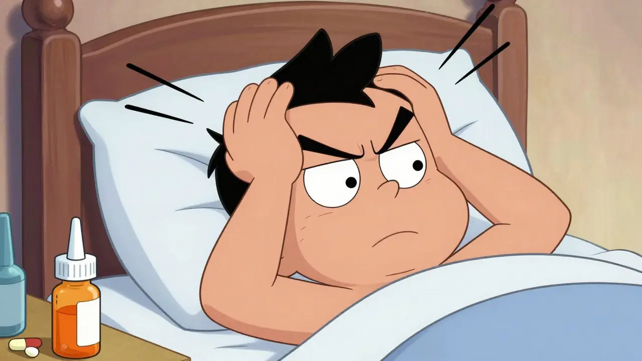
Imagine waking up with a headache so intense it feels like someone is pressing on your eyeballs. Your vision is blurry, you see halos around streetlights, and your eyes look red and swollen. You might think it’s a bad migraine or severe allergies, but if you recently took certain common medications, you could be facing acute angle-closure glaucoma (AACG). This is not just an uncomfortable symptom; it is a true medical emergency where permanent blindness can occur within 24 to 72 hours if untreated.
While many people associate glaucoma with gradual age-related vision loss, drug-induced AACG strikes suddenly. It happens when specific drugs trigger a mechanical blockage in the eye's drainage system, causing intraocular pressure (IOP) to spike dangerously high. Understanding which medications cause this, who is at risk, and how to spot the warning signs early can mean the difference between saving your sight and losing it permanently.
What Is Drug-Induced Acute Angle-Closure Glaucoma?
Acute angle-closure glaucoma is a sudden blockage of the trabecular meshwork resulting in rapid elevation of intraocular pressure. In the context of medication use, it occurs when a drug causes the pupil to dilate or swells the iris tissue, physically blocking the flow of aqueous humor out of the eye. Unlike primary open-angle glaucoma, which develops silently over years, this condition presents with acute, painful symptoms requiring immediate intervention.
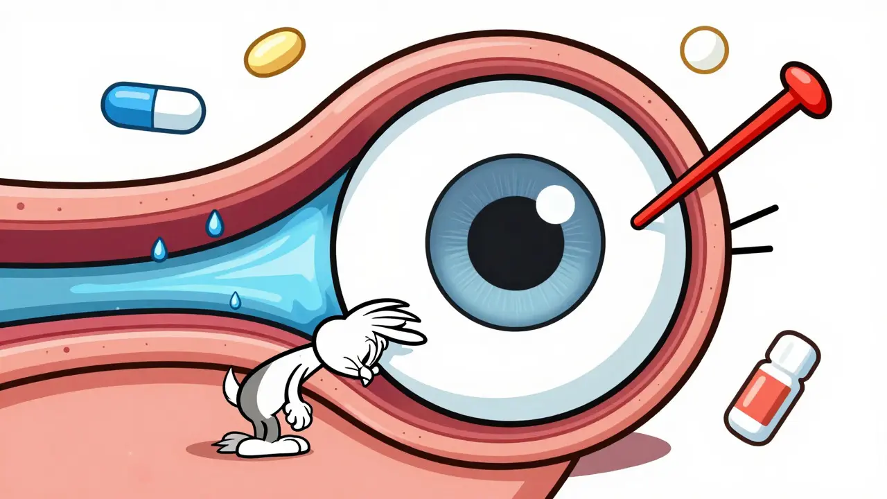
The mechanism is largely mechanical. The most common pathway is pupillary block. When a medication causes the pupil to dilate (mydriasis), the edge of the iris can press against the lens. This creates a seal that prevents fluid from moving from the back of the eye to the front, where it drains. As fluid builds up behind the iris, the iris bows forward, further narrowing the angle and eventually closing it completely. Other mechanisms include plateau iris configuration, where the ciliary processes are positioned anteriorly, and malignant glaucoma, involving swelling of the ciliary body.
This condition accounts for approximately 10-15% of all AACG cases globally. It is a preventable emergency, yet it continues to cause significant vision loss because patients and sometimes healthcare providers fail to recognize the link between recent medication intake and sudden eye pain.
High-Risk Medications to Watch Out For
Not every pill or drop carries this risk, but several common classes of drugs are well-documented triggers. If you have narrow angles in your eyes, these medications require extra caution:
- Adrenergic Agents: Phenylephrine, found in many nasal decongestants and some eye drops, is responsible for about 35% of documented cases. Ephedrine, used in some bronchodilators, also poses a risk.
- Anticholinergics: These block acetylcholine, leading to pupil dilation. Tropicamide (used in eye exams) and atropine are classic examples. Antihistamines like diphenhydramine and tricyclic antidepressants such as amitriptyline also fall into this category.
- Sulfonamides: Sulfa-based drugs, including acetazolamide and topiramate, can cause ciliary body edema, pushing the lens-iris diaphragm forward. Topiramate specifically has a black box warning for this risk.
- Serotonergic Antidepressants: Selective serotonin reuptake inhibitors (SSRIs) like paroxetine comprise about 12% of cases, likely due to their mild anticholinergic properties.
- Antihistamines/Decongestants: Over-the-counter allergy meds containing pseudoephedrine or first-generation antihistamines are frequent culprits in patient reports.
| Medication Class | Common Examples | Primary Mechanism | Estimated % of Cases |
|---|---|---|---|
| Adrenergic Agents | Phenylephrine, Ephedrine | Pupil Dilation (Mydriasis) | ~35% |
| Anticholinergics | Tropicamide, Amitriptyline, Diphenhydramine | Pupil Dilation & Iris Swelling | ~28% |
| Sulfonamides | Topiramate, Acetazolamide | Ciliary Body Edema | ~15% |
| SSRIs | Paroxetine, Fluoxetine | Mild Anticholinergic Effect | ~12% |
Who Is Most At Risk?
Anatomy plays a huge role. Not everyone who takes these medications will develop AACG. The condition primarily affects individuals with anatomically predisposed eyes, specifically those with narrow iridocorneal angles.
Ethnicity is a significant factor. East Asian populations have a 2.2 times higher risk than Caucasian populations due to shallower anterior chambers. Narrow angles are present in approximately 8.5% of Asian populations compared to 3.8% of White populations. Other risk factors include hypermetropia (farsightedness), short axial eye length (less than 22 mm), and older age. Women are also more susceptible than men due to generally smaller eye dimensions.
A critical issue is lack of awareness. Studies show that only 25% of patients know they have narrow angles before an attack occurs. Many people go through life without ever having a gonioscopy (angle examination), leaving them vulnerable to drug-induced attacks without realizing the danger.
Recognizing the Symptoms: A Medical Emergency
The onset of drug-induced AACG is usually abrupt and painful. Patients often describe the experience as one of the worst headaches of their lives. Key symptoms include:
- Severe Eye Pain: Often accompanied by a feeling of pressure or fullness in the eye.
- Headache: Typically unilateral (one-sided) and intense.
- Nausea and Vomiting: Common due to the severe rise in intraocular pressure.
- Visual Disturbances: Blurred vision, halos around lights, and decreased visual acuity.
- Red Eye: Conjunctival injection (redness) and corneal edema (cloudiness).
- Fixed Pupil: The pupil may appear mid-dilated (4-6 mm) and unresponsive to light.
Intraocular pressure typically spikes to 40-80 mm Hg, far above the normal range of 10-21 mm Hg. Irreversible optic nerve damage can begin within 6-12 hours of symptom onset. If IOP remains above 40 mm Hg for more than 24 hours, permanent visual field loss becomes highly likely. Time is of the essence.
Diagnosis and Immediate Treatment
Diagnosis involves measuring intraocular pressure with tonometry and examining the eye structures using gonioscopy or ultrasound biomicroscopy. However, in an emergency setting, treatment often begins immediately based on clinical suspicion to lower pressure quickly.
Standard emergency protocols include:
- Medical Therapy: Administration of pilocarpine 2% eye drops to constrict the pupil and break the block. Intravenous mannitol (1-2 g/kg) may be given to rapidly reduce overall eye pressure.
- Laser Peripheral Iridotomy (LPI): A laser procedure performed within 24 hours to create a small hole in the iris, allowing fluid to bypass the blockage. This is the definitive treatment for pupillary block AACG.
- Discontinuation of Offending Agent: Stopping the medication that triggered the attack is crucial to prevent recurrence.
Interestingly, treatment strategies vary depending on the inciting medication. For example, if the cause is sulfonamide-induced ciliary body edema, simple pupil constriction might not work as effectively as in pupillary block cases, and anti-inflammatory agents may be required. This nuance requires expert ophthalmologic assessment.
Prevention: Screening and Medication Choices
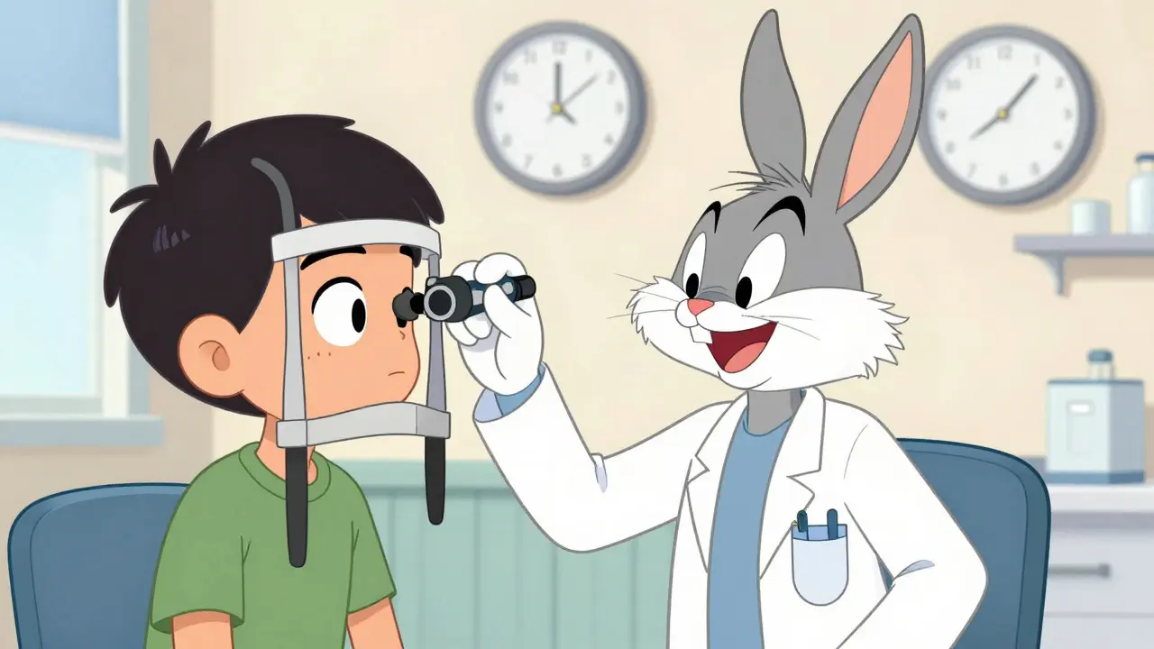
Prevention is possible, but it requires proactive screening. The American Academy of Ophthalmology recommends gonioscopy for all patients over 40 before prescribing high-risk medications. This exam takes only 5-7 minutes per eye and uses a specialized lens to visualize the drainage angle.
If you have narrow angles (Shaffer grading score ≤2), alternatives should be considered. For allergies, loratadine (a non-sedating antihistamine with less anticholinergic effect) is safer than diphenhydramine. For asthma, selective beta-2 agonists like formoterol are preferred over epinephrine. Always inform your doctor about any history of eye problems or narrow angles before starting new prescriptions.
Healthcare systems are improving documentation standards. Electronic health records now increasingly include mandatory glaucoma risk alerts for high-risk prescriptions. However, gaps remain. Only 42% of primary care physicians routinely screen for glaucoma risk before prescribing high-risk medications. Patient education is key-ask your pharmacist or doctor if a new medication could affect your eye pressure.
Frequently Asked Questions
Can over-the-counter cold medicine cause glaucoma?
Yes. Decongestants containing phenylephrine or pseudoephedrine can dilate the pupil and trigger acute angle-closure glaucoma in susceptible individuals. If you have known narrow angles, check with your pharmacist before using these products.
How long does it take to lose vision from untreated AACG?
Irreversible optic nerve damage can start within 6 to 12 hours. Permanent visual field loss is likely if intraocular pressure stays above 40 mm Hg for more than 24 hours. Immediate treatment is critical.
Is drug-induced AACG different from regular glaucoma?
Yes. Regular open-angle glaucoma develops slowly and is often painless. Drug-induced AACG is acute, painful, and caused by a sudden mechanical blockage triggered by medication. It requires emergency intervention rather than long-term management alone.
Who should get a gonioscopy exam?
Ideally, anyone over 40, especially those with farsightedness, Asian heritage, or a family history of glaucoma, should have a baseline gonioscopy. It helps identify narrow angles before high-risk medications are prescribed.
Can LASIK surgery fix angle-closure glaucoma?
No. LASIK corrects refractive errors like nearsightedness. Angle-closure glaucoma is treated with laser peripheral iridotomy (LPI) or surgical procedures to widen the drainage angle. Consult an ophthalmologist for the appropriate treatment plan.