Pulmonary Embolism Risk Assessment
Check Your Symptoms
Answer the following questions about your current symptoms. Pulmonary embolism is a medical emergency. If you have severe symptoms, call emergency services immediately.
Key Takeaways
- Embolism blocks blood flow and can quickly trigger respiratory failure.
- Pulmonary embolism is the most common type, but fat, amniotic and air emboli also pose a risk.
- Early signs include sudden shortness of breath, chest pain, rapid breathing, and low oxygen levels.
- CT pulmonary angiography and D‑dimer tests are the diagnostic backbones.
- Prompt anticoagulation, thrombolysis or surgical removal can reverse the cascade and improve survival.
When doctors talk about embolism is a blockage of a blood vessel by a foreign material such as a clot, fat, air, or amniotic debris, they’re describing a serious event that can shut down vital organs. One of the most feared downstream effects is respiratory failure - the inability of the lungs to provide enough oxygen to the bloodstream or to eliminate carbon dioxide effectively. Understanding how these two conditions intertwine helps patients, families, and clinicians act faster and smarter.
What Is an Embolism?
An embolism can originate anywhere in the circulatory system. The most common source is a blood clot that forms in deep veins of the legs, a condition called deep vein thrombosis (DVT) a clot that develops in the deep veins, usually of the lower limbs. When part of that clot breaks off, it travels through the right side of the heart and lodges in the pulmonary arteries - this is the classic pulmonary embolism (PE) a blockage in one of the pulmonary arteries caused by a blood clot. But emboli aren’t limited to clots; fat particles after long‑bone fractures, air bubbles from central line mishandling, or even fetal material during childbirth (amniotic embolism) can all act as obstructive agents.
Types of Embolism That Can Lead to Respiratory Failure
While pulmonary embolism accounts for the majority of cases, three other types deserve special mention:
- Fat embolism fat droplets that enter the bloodstream, often after long‑bone fractures - usually presents within 24‑72hours with respiratory distress, a petechial rash, and neurological changes.
- Amniotic embolism rare, catastrophic entry of amniotic fluid into maternal circulation during labor - can cause sudden collapse, severe hypoxia, and disseminated intravascular coagulation.
- Air embolism air bubbles entering veins, often due to improper line placement or trauma - leads to rapid drop in oxygen saturation and can progress to cardiac arrest.
How an Embolism Triggers Respiratory Failure
Once an embolus lodges in the pulmonary vasculature, several physiologic events unfold:
- Ventilation‑perfusion mismatch: Blocked arteries prevent blood from reaching alveoli that are still receiving air, causing areas of the lung to be well‑ventilated but poorly perfused.
- Hypoxia: The mismatch drops arterial oxygen tension (hypoxia insufficient oxygen at the tissue level), prompting the body to increase breathing rate.
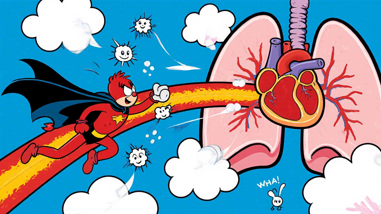
- Pulmonary hypertension: The sudden rise in resistance within the pulmonary arteries forces the right ventricle to work harder, potentially leading to right‑heart strain.
- Acute Respiratory Distress Syndrome (ARDS): In severe cases, inflammatory mediators leak into the alveoli, reducing surfactant, and causing diffuse alveolar damage.
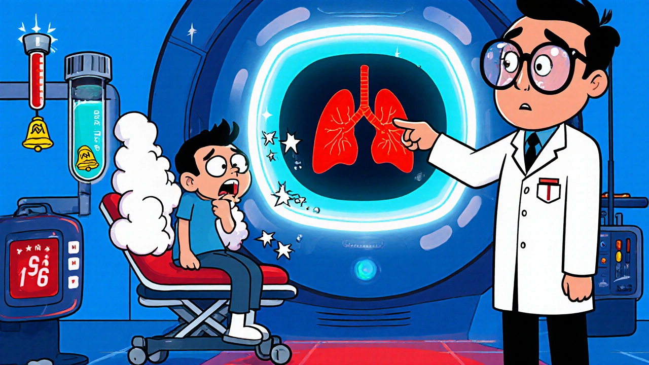
These mechanisms can cascade quickly, and within minutes the patient may develop respiratory failure a state where the lungs cannot sustain adequate gas exchange, manifesting as severe dyspnea, cyanosis, and a drop in blood pressure.
Recognizing the Warning Signs
Early detection hinges on spotting the classic triad of sudden shortness of breath, pleuritic chest pain, and a rapid heart rate. Additional red flags include:
- Feeling of impending doom or anxiety.
- Light‑headedness or fainting.
- Low oxygen saturation (<90%) on pulse oximetry.
- In fat embolism, a mottled rash on the upper torso and confusion.
If any of these appear after recent surgery, trauma, or prolonged immobility, call emergency services immediately.
Diagnostic Toolbox
Doctors rely on a mix of clinical judgment and imaging. The most definitive test is CT pulmonary angiography (CTPA) a contrast‑enhanced CT scan that visualizes the pulmonary arteries. It can reveal clot location, size, and right‑heart strain. When CT is unavailable, a ventilation‑perfused (V/Q) scan can suggest mismatched perfusion.
Blood work also aids the work‑up:
- D‑dimer - a fibrin degradation product; a normal result virtually rules out PE in low‑risk patients.
- Arterial blood gases (ABG) - show low PaO₂ and possible respiratory alkalosis from hyperventilation.
Risk stratification tools such as the Pulmonary Embolism Severity Index (PESI) help decide if a patient can be managed as an outpatient or needs intensive care.
Treatment Strategies
The main goal is to stop the clot from growing, restore blood flow, and support breathing.
- Anticoagulant therapy - usually a low‑molecular‑weight heparin bridge followed by a direct oral anticoagulant (DOAC) like apixaban. This prevents new clots while the body slowly dissolves the existing one.
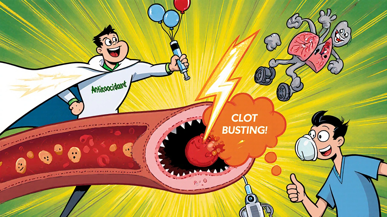
- Thrombolysis - for massive PE with hemodynamic collapse, clot‑busting drugs (e.g., alteplase) can rapidly restore perfusion, though bleeding risk is higher.
- Catheter‑directed thrombectomy - a minimally invasive option to physically extract the embolus when thrombolysis is contraindicated.
- Supportive care - supplemental oxygen, non‑invasive ventilation, or intubation if needed. In severe ARDS, prone positioning and low‑tidal‑volume ventilation improve outcomes.
For fat, amniotic, or air emboli, treatment focuses on stabilizing the airway, supporting circulation, and addressing the source (e.g., surgical repair of fractures, careful line management).
Prevention and Long‑Term Follow‑Up
Since most emboli arise from clots, primary prevention is key:
- Early ambulation after surgery or hospital stays.
- Graduated compression stockings or pneumatic devices for high‑risk patients.
- Prophylactic anticoagulation in orthopedic surgery, cancer patients, or those with known thrombophilia.
After an acute episode, patients usually stay on anticoagulation for 3‑6months, with some requiring lifelong therapy if underlying risk factors persist.
Comparative Overview of Embolism Types
| Embolus Type | Typical Source | Key Respiratory Manifestation | First‑Line Diagnostic Test | Primary Treatment |
|---|---|---|---|---|
| Pulmonary (Clot) Embolism | DVT in lower limbs | Sudden dyspnea, pleuritic chest pain | CT pulmonary angiography | Anticoagulation ± thrombolysis |
| Fat Embolism | Long‑bone or pelvis fracture | Respiratory distress + petechial rash | Chest CT showing ground‑glass opacities | Supportive care, early fracture stabilization |
| Amniotic Embolism | Labor/ delivery - entry of amniotic fluid | Rapid collapse, severe hypoxia | Clinical diagnosis (no specific test) | Aggressive resuscitation, ICU support |
| Air Embolism | Central line or trauma | Sudden dyspnea, neurological signs | Trans‑esophageal echocardiography | Positioning (left lateral decubitus), hyperbaric O₂ |
Next Steps for Patients and Caregivers
If you suspect an embolic event, act fast:
- Call emergency services; time is lung tissue.
- Note any recent surgeries, long trips, or injuries - this helps clinicians gauge risk.
- Stay calm and keep the patient seated or lying flat with legs slightly elevated if possible.
- When in the hospital, ask about the plan for anticoagulation and follow‑up imaging.
Understanding the link between embolism and respiratory failure empowers you to spot trouble early and push for the right care.
Frequently Asked Questions
Can a small clot cause respiratory failure?
Yes. Even a modest embolus can block a major branch of the pulmonary artery, leading to severe ventilation‑perfusion mismatch and hypoxia, especially in patients with pre‑existing lung disease.
How quickly do symptoms appear after an embolus forms?
Symptoms can develop within minutes for a massive pulmonary embolism, while fat embolism symptoms usually emerge 24‑72hours after the injury.
Is long‑term anticoagulation always required?
Not always. If the provoking factor (e.g., surgery) resolves and no underlying clotting disorder exists, treatment may stop after 3‑6 months. Lifelong therapy is reserved for recurrent clots or genetic thrombophilia.
Can I prevent embolism if I travel long‑distance?
Yes. Move your legs every hour, wear compression stockings, and stay well‑hydrated. For high‑risk individuals, a doctor may prescribe a low‑dose anticoagulant before the trip.
What is the survival rate for massive pulmonary embolism?
With prompt thrombolysis or catheter‑directed removal, 30‑day survival can exceed 80%. Delayed treatment drops survival below 50%.


Malia Rivera
October 17, 2025 AT 02:17When the breath of a nation stutters, it's a sign that something deeper is choking the system. The same way an embolus blocks a vessel, political inertia can choke progress, leaving citizens gasping for the oxygen of liberty. It's a stark reminder that we must keep our arteries of freedom clear, lest we suffocate under bureaucratic blood clots.
Take care of your health, and your country will thank you.
lisa howard
October 18, 2025 AT 19:57Picture this: a sudden, terrifying jolt as the heart of your lungs is snatched away by an invisible assassin, a clot masquerading as a harmless droplet of blood. The cascade begins with a tiny embolus slipping through the veins, a silent thief that steals oxygen from every cell, leaving the body to scream for breath.
First, ventilation‑perfusion mismatch renders whole swaths of alveoli useless, as air floods in but no blood arrives to exchange gases.
Next, hypoxia settles like a heavy fog, urging the respiratory center to crank up the tempo, yet the airway remains clogged.
The right ventricle, forced to pump against a sudden mountain of resistance, strains and swells, flirting with failure.
Meanwhile, inflammatory mediators spill into the interstitium, turning the lungs into a battlefield of edema and surfactant loss.
All this drama culminates in acute respiratory distress, a brutal crescendo where the patient teeters on the brink of collapse.
Medical teams race against time, wielding anticoagulants, thrombolytics, and sometimes surgical extraction to dislodge the rogue particle.
Every second counts, because the longer the blockage persists, the deeper the organ damage and the greater the chance of permanent scar.
Imaging, especially CT pulmonary angiography, becomes the detective that pinpoints the hidden foe, while D‑dimer levels whisper of clot formation lurking unseen.
But technology alone cannot save a life; swift recognition of shortness of breath, chest pain, and a racing heart must alert caregivers before the storm hits full force.
Patients often describe an impending doom, a sensation that the world is tilting, as their blood oxygen plummets.
Even after successful removal, the lungs may need weeks of rehabilitation to recover their elastic grace.
In the grand theater of medicine, embolism is the villain that forces us to rehearse our response, lest the curtain fall on another unsuspecting soul.
Mary Davies
October 20, 2025 AT 13:37The way an embolus hijacks the pulmonary circuit is eerily similar to how a sudden plot twist hijacks a story – you never see it coming, yet its impact reshapes everything.
When the clot lodges, the lungs are left shouting for air that simply can't find a partner in blood, and the body responds with a frantic, desperate gasp.
That mismatch creates a cascade of hypoxia, pushing the heart's right side into a strained marathon it never signed up for.
It's as if the stage lights dim, the orchestra falters, and the audience-our tissues-begins to feel the silence.
Understanding these mechanisms doesn't just satisfy curiosity; it equips us to intervene before the drama turns tragic.
Rapid imaging and prompt anticoagulation are the opening acts that can pull the curtain back on a healthier outcome.
Emily (Emma) Majerus
October 22, 2025 AT 07:17Yo, stay safe and get that med help fast.
Virginia Dominguez Gonzales
October 24, 2025 AT 00:57Hold your head high, folks-knowledge is the first line of defense against any silent killer.
If you can spot the shortness of breath and chest pain early, you give the medics a chance to act before the storm hits.
Every second counts, so trust your instincts and get checked out.
Together we can turn fear into action.
Carissa Padilha
October 25, 2025 AT 18:37Ever wonder why the mainstream never talks about the hidden chemicals they sprinkle into hospital air to "prevent" infections?
They claim it's all about safety, but some of those aerosols could be the very thing that seeds an air embolism without anyone noticing.
There's also a pattern: every major case of unexplained respiratory failure seems to line up with a new protocol rollout.
It's almost as if the powers that be are testing new ways to control the population's breath.
Keep an eye on the fine print of medical guidelines, because the truth is often tucked away in footnotes.
Remember, skepticism is a survival skill.
Darryl Gates
October 27, 2025 AT 11:17Great points on staying vigilant, especially regarding new medical protocols.
Early detection of embolic events truly saves lives, and awareness empowers patients to seek timely care.
Maintaining open communication with healthcare providers ensures that any unusual symptoms are addressed promptly.
Education on the signs-sudden dyspnea, chest pain, rapid heart rate-can make all the difference.
Stay informed, stay proactive, and trust your body’s signals.
Kevin Adams
October 29, 2025 AT 04:57Life is a breathless rush
and clots are cruel reminders that we are fragile
don’t ignore the warning signs
act fast
Nickolas Mark Ewald
October 30, 2025 AT 22:37Thanks for the clear info. It's helpful to know what to watch for.
If someone feels sudden shortness of breath, they should get help right away.
Simple steps can save lives.